 Bloggers Trend Keeping You Up To Date
The human neck has seven cervical vertebrae, all separated by shock absorbers: the intervertebral discs.
A herniated cervical disc is an injury to a disc that separates two vertebrae in your neck.
The lesion of the disc (disc disease), caused either by trauma or more often by wear and tear, can cause compression on the nerve structures.
The cervical disc herniation sits at the back of the disc and can then compress either the nerve exiting between the two vertebrae (the nerve root) or even if the herniation is larger, compress the entire spinal cord which is located behind the disk.
When this compression acts on the outgoing nerve (root), it causes pain in the arm called cervicobrachial neuralgia, sometimes reaching the fingers.
The painful path provides information on the level of the affected disc.
Evolution of cervical disc herniation
The more the compression is important and lasting the more the risk of serious lesions increases, the initial pain can then intensify, then tingling and loss of sensitivity appear, or even problems with loss of strength in the hand or even the arm.
The pain is usually made worse by movement of the head and neck.
When the hernia is very large, and located in the middle of the disc, it can directly compress the main nerve structure, the spinal cord. This can result in a feeling of weakness in the legs (limiting your walking range) and dropping objects.
The spontaneous evolution can in certain cases lead to an improvement or disappearance of neuralgia pain, but we can only consider patient in the absence of deficient neurological disorders and if the pain remains bearable.
Paraclinical examinations for herniated cervical disc
Even if the surgeon examining you may already have a precise idea of the disc level causing the problem, a number of additional examinations are now useful, to confirm the exact location of the lesion but also to eliminate a number of diseases, which could give misleading signs.
These are the following exams.
Standard radiography
It makes it possible to recognize the level of the disc reached, because in general, the disc is less high and partially overwritten. Most often, hernias are located between the 5th and 6th cervical vertebrae and / or between the 6th and 7th cervical vertebrae.
The x-ray will also look for signs of osteoarthritis in older lesions, and also study the general curvature of the cervical spine. This radiological examination does not present any danger of irradiation, unless it was repeated very often.
It is sometimes useful to order radiological examinations in the flexion and extension position to assess the flexibility of the column and the instability of the affected disc.
The scanner (or computed tomography)
The scanner also uses X-rays and allows the disc to be viewed more precisely, in slices.
It makes it possible to clearly specify the size of the hernia, its position and whether it is accompanied by bone constructions linked to osteoarthritis. Sometimes the radiologist decides to give an intravenous injection of iodine, in order to better see the environment of the hernia. You should know that the scanner uses a much larger quantity of X-rays than the simple radiography.
Magnetic resonance imaging (MRI)
It allows to give additional information to that of the scanner, by showing better the surrounding soft tissues and by making it possible to give information on the quality of the tissues, it shows on the other hand the bone structures less well.
It is particularly good at detecting lesions of the spinal cord (cervical myelopathy) in cases of narrowing of the duct due to osteoarthritis or a very large hernia.
Electromyogram (EMG)
It allows to study the quality of functioning of the nerves, and to confirm and specify the severity of the nerve compression.
More complex exams
More complex examinations (motor and sensory evoked potentials) are sometimes requested in order to record the functioning of the spinal cord.
These examinations make it possible to account for the severity of the nerve damage, which sometimes explains the differences in recovery.
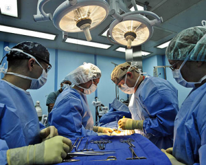
In more complex cases, the patient can be referred to hernia surgery instrument set.
For more details, please visit: jimymedical.co.uk