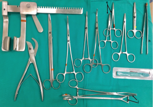
Veterinary cardiology is a discipline that has benefited from recent advances in human cardiology and its own research. This is especially true for the diagnosis of heart disease with the development of echocardiography, electrocardiography and with the help of open heart surgical instrument set and Holter.
Medical treatment has also made many advances, with the proof of the effectiveness of several classes of drugs such as angiotensin converting enzyme inhibitors, inodilators or latest generation diuretics, making it possible to improve the quality of life and extend the life of animal heart.
Certain animal heart conditions may also benefit from curative surgery. However, this remains limited to extra-cardiac interventions (without opening the heart) carried out in veterinary clinics: ligation of the ductus arteriosus, excision of tumors at the base of the heart or of the pericardium (pericardectomy).
There is also the possibility of placing a pacemaker, the purpose of which is to treat animals with severe arrhythmias. There is no specific pacemaker designed for dogs. However, human pacemakers are adaptable to animals.
In recent years, other more complex surgical corrections, because “intra-cardiac”, are available to domestic carnivores. They are reserved for university centers or research centers with a large technical platform, equipped with high-tech equipment, allowing two surgical approaches: one by endovascular route (so-called “interventional” surgery performed without opening the heart) and the other open heart requiring the establishment of extracorporeal circulation (ECB).
The partnership between the Alfort Cardiology Unit (UCA) and cardiac surgeons from IMM research (UCA-IMMR), created more than 20 years ago at the initiative of professors JL Pouchelon, V Chetboul and F Laborde, made it possible to operate and save animals suffering from various heart diseases, most often congenital, by one or the other of these two approaches.
General: Interventional catheterization was recognized as an innovative and useful technique following the work of French physiologist André Cournan whose research on this subject earned him the award of the Nobel Prize for medicine in 1956. This technique consists, under the supervision of an image intensifier, by inserting a catheter into a vein or artery to access the heart.
Since the material is introduced into the vessels through the skin, these are also referred to as endoluminal or percutaneous procedures. These endovascular procedures thus make it possible, nowadays, to avoid certain open-heart surgeries, in particular in pulmonary stenosis (narrowing of the pulmonary artery) and, more recently, in the persistence of the ductus arteriosus in domestic carnivores.
Persistence of the ductus arteriosus: the persistence of the ductus arteriosus is one of the most common congenital heart diseases in dogs. The ductus arteriosus, present in the fetus, closes within the first 72 hours of life. The persistence of the ductus arteriosus is a congenital malformation characterized by the absence of closure of this duct, thus causing a mixture of blood between the aorta and the pulmonary artery with often deleterious effects (40% survival at 1 year without surgical correction).
The closure of the ductus arteriosus is conventionally performed by thoracotomy (opening of the thorax) and ligation of the duct after its dissection.
These endovascular devices specifically designed for dogs were introduced in the late 2000s and have the advantage of being specifically adapted to the shape and size of the arterial ducts in this species. The risk of embolization is therefore particularly reduced.
This technique presents, moreover, very good results with many advantages: short operating time limiting the anesthetic risks, absence of dissection of the canal thus reducing the hemorrhagic risks, and absence of opening of the thorax allowing a short postoperative hospitalization duration.
Pulmonary stenosis: Similarly, balloon dilation (or percutaneous pulmonary angioplasty) is the technique of choice for correcting – without opening the heart – another very common congenital malformation of dogs, pulmonary stenosis, representing up to 32% of Congenital heart disease in this species, French and English Bulldogs being the most predisposed breeds. The technique involves passing a balloon through catheters previously introduced into a vein to the place of the arterial obstacle.
For more details, please visit: jimymedical.co.uk
 Bloggers Trend Keeping You Up To Date


